
Home » Equine Lameness » Scientific Background
During the dynamic part of the lameness examination, most information about the presence of lameness and the affected leg is gathered during trot. Trot is a symmetrical two-beat gait with limbs moving in diagonal pairs (Hildebrand, 1965) in near-synchrony. Assessment at trot takes advantage of the increased forces being exerted on the limbs (Merkens et al., 1986; Merkens et al., 1993) due to the increased speed and bouncing gait (Biewener, 2003). A painful focus in the limb can thus be exacerbated, leaving the horse little option to redistribute weight unless adopting an asymmetrical movement pattern. The resulting movement asymmetry, which is visually assessed by clinicians, is the main indicator of lameness.
Both kinematic (based on movement) and kinetic (based on forces) approaches have yielded sensitive measures for objective lameness quantification during trot on the straight line. Some of the most reliable objective features to detect lameness are based on upper body movement. Other very reliable indicators are differences in fetlock hyperextension or differences in peak vertical ground reaction force and vertical impulse compared between contralateral limbs (Buchner et al., 1996b; Ishihara et al., 2005; Weishaupt et al., 2006; Weishaupt, 2008).
Upper body movement lends itself to clinical lameness assessment, as changes are – within the bounds of perceptual limitations – readily visible to the eye. Studies into head and trunk movement have investigated the response of several upper-body landmarks to lameness. Investigations concerned head, withers, os sacrum and tubera coxae movement adaptations (Buchner et al., 1996a), selected locations such as only head or only the pelvis (Peham et al., 1996; Peham et al., 1999; Keegan et al., 2000; Kramer et al., 2000; Keegan et al., 2001; Peham et al., 2001; Keegan et al., 2003; Kramer et al., 2004; Pfau et al., 2007), locations distributed along the back (Audigie et al., 2002) and centre of mass movement adaptations (Buchner et al., 2001; Halling Thomsen et al., 2010). Alternatively, movement of the sternum has been measured (Barrey et al., 1994; Barrey and Desbrosse, 1996).
These studies found that asymmetry in vertical acceleration, velocity and / or displacement of most upper body landmarks reflected lameness well (Buchner et al., 1996a; Peham et al., 1996; Uhlir et al., 1997; Church et al., 2009). Today, vertical head movement asymmetry is considered the most reliable kinematic lameness pointer for forelimb lameness (Peloso et al., 1993; Buchner et al., 1996a; Keegan et al., 1997; Peham et al., 1999; Keegan et al., 2000; Keegan et al., 2001; Keegan et al., 2003; Keegan et al., 2004) while vertical movement asymmetry of os sacrum and tubera coxae is the most reliable kinematic pointer for hind limb lameness (May and Wyn-Jones, 1987; Buchner et al., 1993; Buchner et al., 1996a; Kramer et al., 2000; Peham et al., 2001; Kramer et al., 2004; Pfau et al., 2007; Church et al., 2009).
Head movement can be used to detect forelimb lameness (Buchner et al., 1996a; Keegan et al., 2000; Keegan et al., 2001; Keegan et al., 2003). In a sound horse, vertical head displacement resembles a symmetrical, double-sinusoidal pattern (Buchner et al., 1993; Peham et al., 1999).
With the onset of lameness, this pattern is disturbed (Peloso et al., 1993; Buchner et al., 1996a; Peham et al., 1996; Keegan et al., 1997; Peham et al., 1999; Peham et al., 2000), causing asymmetry between the two vertical excursions of the head during successive steps. A modelling study showed that head movement adaptations during forelimb lameness can actually account for the majority of limb unloading (Vorstenbosch et al., 1997). While this results in a straight forward indication of lameness, the pattern of head movement during lameness varies.
For most forelimb lame horses, the head reaches its lowest position near mid-stance of the sound limb. Less commonly, horses for example show increased head elevation following stance of the lame limb while maintaining similar minimum positions, also characterised as ‘push-off lameness’ (Kramer and Keegan, 2004).
In moderate to severe cases of forelimb lameness, head movement changes to a single sinusoidal excursion per stride (Clayton, 1987; Peloso et al., 1993; Christovão et al., 2007). This reduces the movement frequency by 50 % and likely contributes to the perceived ease of determining forelimb lameness.
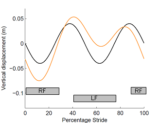
Figure 1: Vertical head movement in a sound horse (black line) and a lame horse (orange line). RF – stance of right forelimb; LF – stance of left forelimb.
In a sound horse, movement is symmetrical: the two maxima reach the same height and the same holds true for the two minima. In a lame horse, you can see that the movement pattern is asymmetrical: the horse is ‘nodding down’ during stance of the right fore, hence it is left forelimb lame.
Pelvis movement can be used to detect hind limb lameness (May and Wyn-Jones, 1987; Buchner et al., 1993; Buchner et al., 1996a; Kramer et al., 2000; Peham et al., 2001; Kramer et al., 2004; Pfau et al., 2007; Church et al., 2009).
This gets a bit more tricky than forelimb lameness signs, since the presence of not only translation but also rotation of the whole pelvis results in different movement of mid-pelvis (os sacrum) and of the hips (tubera coxae): axial rotation of the pelvis causes asymmetry within the left and right tuber coxae movement pattern even in sound horses (Buchner et al., 1993; Buchner et al., 1996a; Kramer et al., 2000; Church et al., 2009). Tubera coxae asymmetry varies notably between but little within horses, indicating different magnitudes of pelvic rotation between horses (Buchner et al., 1996a). It remains unclear whether lame horses show systematic changes in pelvic rotation; there are descriptions of rotation away from the lame limb (Weishaupt, 2008), exaggerated rotation during stance of the sound limb (Buchner et al., 1996a) and a rotation offset towards the non-lame side in the absence of an overall change in rotational range of movement (Gomez-Alvarez et al., 2008). In this game, the axial rotation pattern remains unchanged.
Asymmetry in vertical mid-pelvis (respectively os sacrum) movement is a reliable lameness indicator (Buchner et al., 1996a; Kramer et al., 2000; Peham et al., 2001; Audigie et al., 2002; Kramer et al., 2004), similar to head movement. In most cases of hind limb lameness, the os sacrum shows increased upward movement before foot contact of the lame limb (Keegan, 2011), with a variety of movement patterns; these can be described by the phase difference between symmetrical and asymmetrical signal component (Audigie et al., 2002).
When assessing lameness based on comparative hips (respectively tubera coxae) movement, left and right tubera coxae have to be compared: the vertical displacement amplitude for the tubera coxae on the lame side is greater than the subsequent amplitude of the contralateral tubera coxae (May and Wyn-Jones, 1987), often termed a ‘hiking’ movement. While the tuber coxae on the lame side does not consistently rise above the vertical height of the tuber coxae on the sound side, it may consistently drop below it (May and Wyn-Jones, 1987). The tuber coxae on the lame side shows increased upward movement before foot contact of the lame limb.
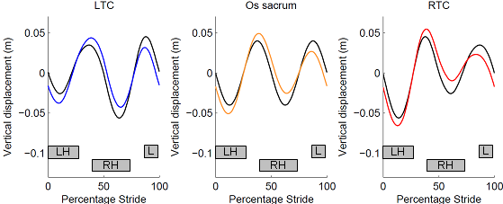
Figure 2: Vertical pelvis movement in a sound horse (black line) and a lame horse (coloured line) based on three landmarks: the mid-pelvis (os sacrum) and hips (LTC – left tubera coxae / left hip; RTC – right tubera coxae / right hip). LH – stance of left hind limb; RH – stance of right hind limb.
In a sound horse, movement of the mid-pelvis is symmetrical in the same way as described for the head. However, rotation of the pelvis causes hips movement to be asymmetrical per se. In a lame horse, you can see that the movement pattern of the mid-pelvis is now asymmetrical and that the pattern of both hips changes; in fact, the left hip is now becoming almost symmetrical!
To detect hind limb lameness, people commonly look for a ‘hip hike’ – an increased upward movement prior to touch-down of the lame limb. You can see that this is reflected in both, the mid-pelvis movement as well as when comparing movement amplitudes of the left and right hip. The horse in this example is right hind limb lame.