
Home » Equine Lameness » Clinical Lameness Examinations
During the lameness examination, the veterinarian has to establish firstly the presence or absence of lameness, secondly the affected limb, thirdly the location of pain and fourthly the cause of the pain. While steps one to three strongly rely on observational skills, step four requires extensive veterinary knowledge of possible diseases. However, you can see that if one gets step one and two wrong, there is not much hope diagnosing the horse correctly. This makes it so important to develop the visual skills that let you decide on the affected limb(s) reliably. Step three and four are the costly and time consuming ones that rely on your ability to get step one and two right.
A lameness examination commonly consists of the following stages after initial inspection of the horse:
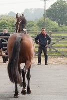
1. Examination on the straight line: After briefly examining the movement pattern at walk, the horse is assessed for movement asymmetry during trot away from and towards the observer; some veterinarians also inspect the horse from the side, as this allows a better appreciation of limb movement, particularly joint flexion and extension.
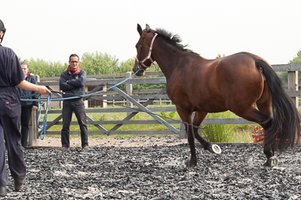
2. Examination during trot in a circle, both on a hard and soft surface: Circling is used to exacerbate lameness and allows the detection of bilaterally symmetrical conditions that may not be obvious during assessment on the straight line. A horse that has similar pain in each forelimb or hindlimb will not be able to preferentially unload a specific lame limb so it may appear to move symmetrically (Miller, 1925, Stashak, 2002, Ross, 2011, Buchner 1993). While horses commonly show a more pronounced response to lunging on a hard compared to a soft surface, for certain conditions the reverse can hold true (Ross, 2011).
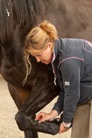
3. Examination after provocation tests such as flexion tests: Flexion tests are believed to exacerbate pain and consequently movement asymmetry, thus helping to identify the affected limb. However, since flexion tests result in a high number of ‘false positives’ (Keg et al., 1997a, Keg et al., 1997b, Ramey, 1997, Verschooten and Verbeeck, 1997, Busschers and Van Weeren, 2001, Armentrout et al., 2011), they are controversial and regarded with caution by most veterinarians.
4. Manual palpation: Palpation is performed at rest to inspect the horse for visible / palpable abnormalities (swelling, pain response etc.) that may allow identification of the affected region and a preliminary diagnosis. However, palpation does not always reveal detectable abnormalities. Equipment such as hoof testers may also be used.
5. Intrasynovial or perineural analgesia: Local analgesic techniques, commonly referred to as ‘nerve blocks’, are performed in order to localise the lameness within the affected limb. These blocks ‘numb’ specific regions in the horse’s limb; they work based on the principle that once the area which is in pain is anaesthetised, the horse will return to its normal symmetrical movement pattern. These procedures help narrow down the affected region in the limb allowing subsequent diagnostic imaging to be appropriately targeted.
6. Diagnostic imaging: Once the lameness has been localised to a specific region, the region will be investigated using diagnostic imaging (X-ray, Scintigraphy, MRI, CT scans etc) in order to precisely establish the cause of lameness.
Once a diagnosis is made, treatment options and the associated prognosis for recovery can be worked out.
Evaluation of the horse at trot on the straight line / circle and interpreting the effect of nerve blocks on changes in baseline lameness is commonly considered the most important skill needed to perform a lameness examination on complex cases and “essential for accurate diagnosis” (Ross, 2011a). If the veterinarian is fails to follow a logical diagnostic pathway, the consequences may include misdiagnosis, misdirected treatment, wasted resources and conflicting professional advice, with prolonged lameness as the consequence for the unfortunate horse.
Hence, a key aspect of a successful lameness examination is the accuracy of visual asymmetry assessment during movement, followed by each of the other stages as appropriate. This visual assessment becomes less reliable in cases where lameness is subtle: movement asymmetry becomes very small and is difficult to see. Hence, research has found disagreement even amongst experienced veterinarians on whether lameness is present and which limb(s) are affected (Pleasant et al., 1997, Keegan et al., 1998, Fuller et al., 2006, Hewetson et al., 2006, Keegan et al., 2010, Starke et al. 2013). This means that different veterinarians may further investigate a different limb of a horse, or send the horse home because they do not consider it observably lame.
Armentrout, A. R., Beard, W. L., White, B. J., Lillich, J. D., 2011. A comparative study of proximal hindlimb flexion in horses: 5 versus 60 seconds. Equine Vet J 44, 420-424.
Busschers, E., Van Weeren, P. R., 2001. Use of the Flexion Test of the Distal Forelimb in the Sound Horse: Repeatability and Effect of Age, Gender, Weight, Height and Fetlock Joint Range of Motion. Journal of Veterinary Medicine Series A 48, 413-427.
Fuller, C. J., Bladon, B. M., Driver, A. J., Barr, A. R. S., 2006. The intra- and inter-assessor reliability of measurement of functional outcome by lameness scoring in horses. The Veterinary Journal, 171, 281-286.
Goubaux, A., Barrier, G. 1892. The exterior of the horse, London, J. B. Lippincott Company.
Hewetson, M., Christley, R. M., Hunt, I. D., Voute, L. C., 2006. Investigations of the reliability of observational gait analysis for the assessment of lameness in horses. Vet Rec, 158, 852-7.
Keegan, K. G., Wilson, D. A., Wilson, D. J., Smith, B., Gaughan, E. M., Pleasant, R. S., Lillich, J. D., Kramer, J., Howard, R. D., Bacon-Miller, C., Davis, E. G., May, K. A., Cheramie, H. S., Valentino, W. L., Van Harreveld, P. D., 1998. Evaluation of mild lameness in horses trotting on a treadmill by clinicians and interns or residents and correlation of their assessments with kinematic gait analysis. American Journal of Veterinary Research, 59, 1370-1377.
Keegan, K. G., Dent, E. V., Wilson, D. A., Janicek, J., Kramer, J., Lacarrubba, A., Walsh, D. M., Cassells, M. W., Esther, T. M., Schiltz, P., Frees, K. E., Wilhite, C. L., Clark, J. M., Pollitt, C. C., Shaw, R., Norris, T., 2010. Repeatability of subjective evaluation of lameness in horses. Equine Vet J, 42, 92-7.
Keg, P. R., Van Weeren, P. R., Back, W., Barneveld, A., 1997a. Influence of the force applied and its period of application on the outcome of the flexion test of the distal forelimb of the horse. Vet Rec 141, 463-6.
Keg, P. R., Van Weeren, P. R., Barneveld, A., Schamhardt, H. G., 1997b. Variations in the force applied to flexion tests of the distal limb of horses. Vet Rec. 141, 435-438.
Miller, W. C., 1925. The physiology of lameness. The Veterinary Record 5, 84-87.
Pleasant, R. S., Moll, H. D., Ley, W. B., Lessard, P., Warnick, L. D., 1997. Intra-articular anesthesia of the distal interphalangeal joint alleviates lameness associated with the navicular bursa in horses. Vet Surg, 26, 137-40.
Ramey, D. W., 1997. Prospective evaluation of forelimb flexion tests in practice: clinical response, radiographic correlations, and predictive value for future lameness. Proceedings of the annual convention of the AAEP 43, 116-120.
Ross, M. W. 2011a. Chapter 2. Lameness in Horses: Basic Facts Before Starting. In: Ross, M. W. & Dyson, S. J. (eds.) Diagnosis and Management of Lameness in Horses. St. Louis, Missouri: Elsevier Saunders.
Ross, M. W. 2011b. Chapter 7. Movement. In: Ross, M. W. & Dyson, S. J. (eds.) Diagnosis and Management of Lameness in Horses. Elsevier Saunders.
Ross, M. W., Dyson, S. J. 2011. Diagnosis and Management of Lameness in Horses. , St. Louis, Missouri, Elsevier Saunders.
Starke, S. D., Raistrick, K. J., May, S. A., Pfau, T., 2013. The effect of trotting speed on the evaluation of subtle lameness in horses. The Veterinary Journal, 197, 245–252.
Stashak, T. S. 2002. Chapter 3. Examination for lameness. In: Stashak, T. S. (ed.) Adams’ Lameness in Horses. Baltimore: Lippincott Williams & Wilkins.
Stashak, T. S., Hill, C. 1996. Practical Guide to Lameness in Horses, Wiley-Blackwell.
Verschooten, F., Verbeeck, J., 1997. Flexion test of the metacarpophalangeal and interphalangeal joints and flexion angle of the metacarpophalangeal joint in sound horses. Equine Veterinary Journal 29, 50-54.
Wyn-Jones, G. 1988. Chapter 1. The Diagnosis of the Causes of Lameness. In: Wyn-Jones, G. (ed.) Equine Lameness. Oxford: Blackwell Scientific Publications.